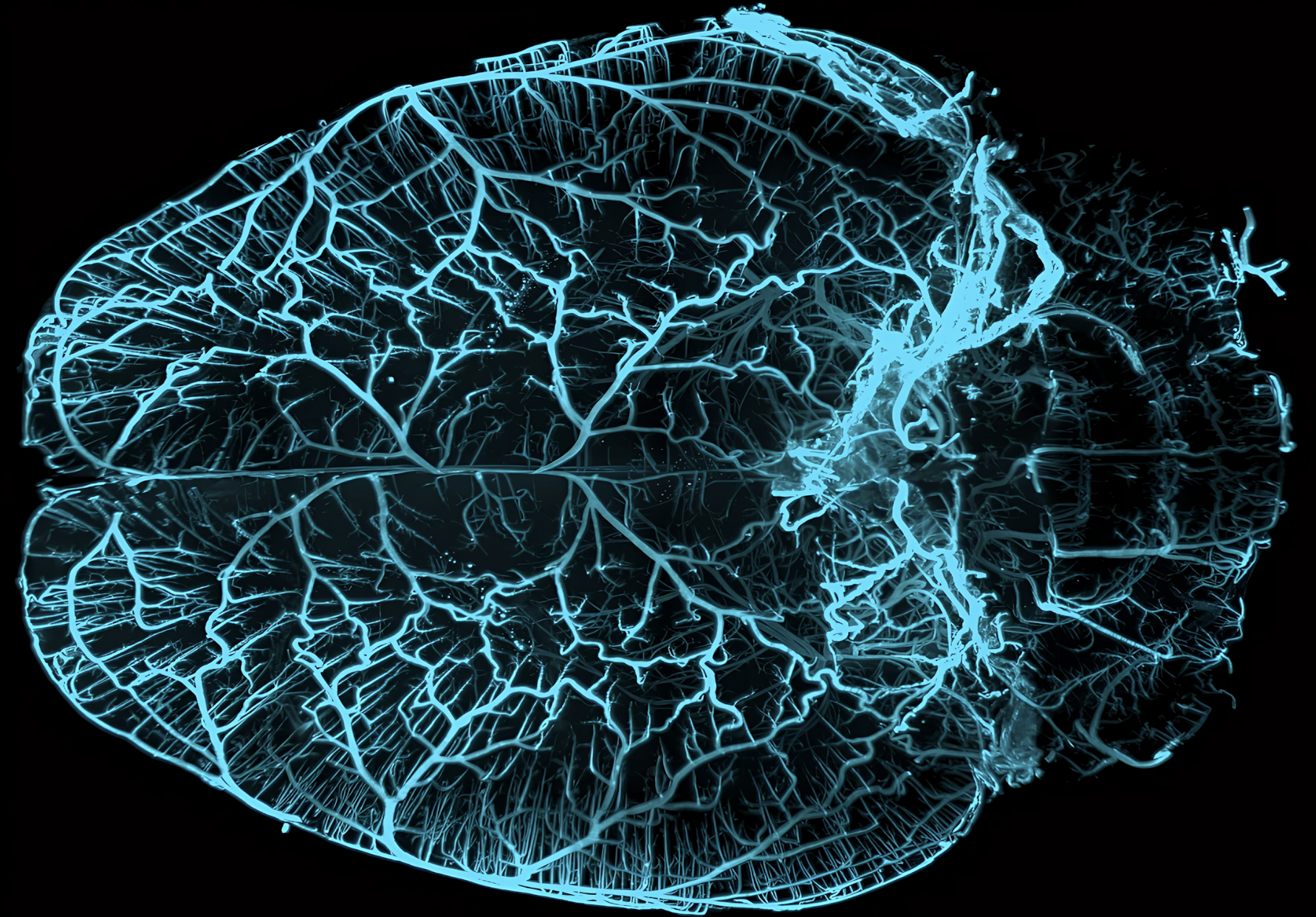
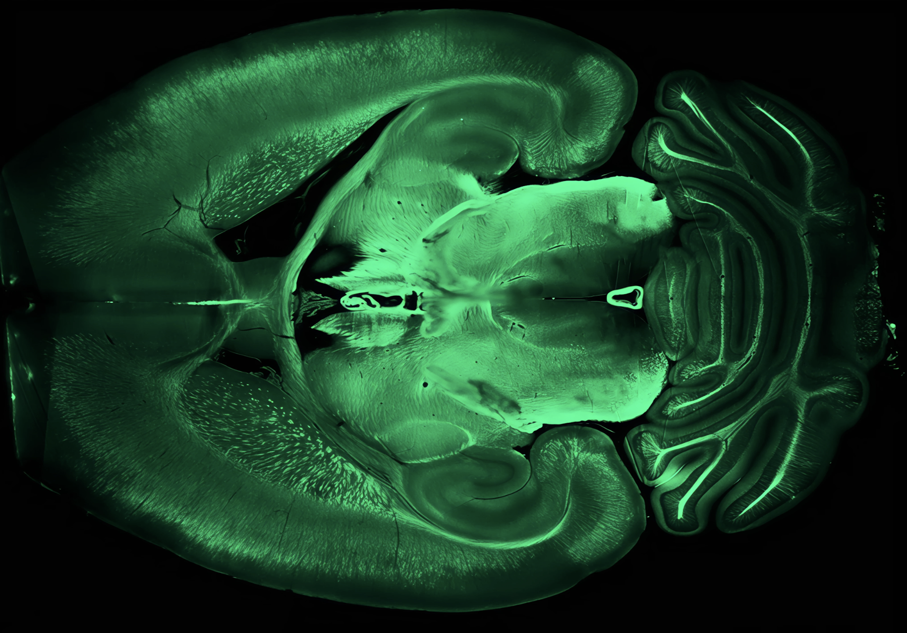
Institut du Cerveau
Myriam Spajer
Myriam Sapjer reveals the vascular network of the brain using Light Sheet microscopy. Each vessel traces a vital path, whose alteration can affect development, balance... and even thought.
AVC
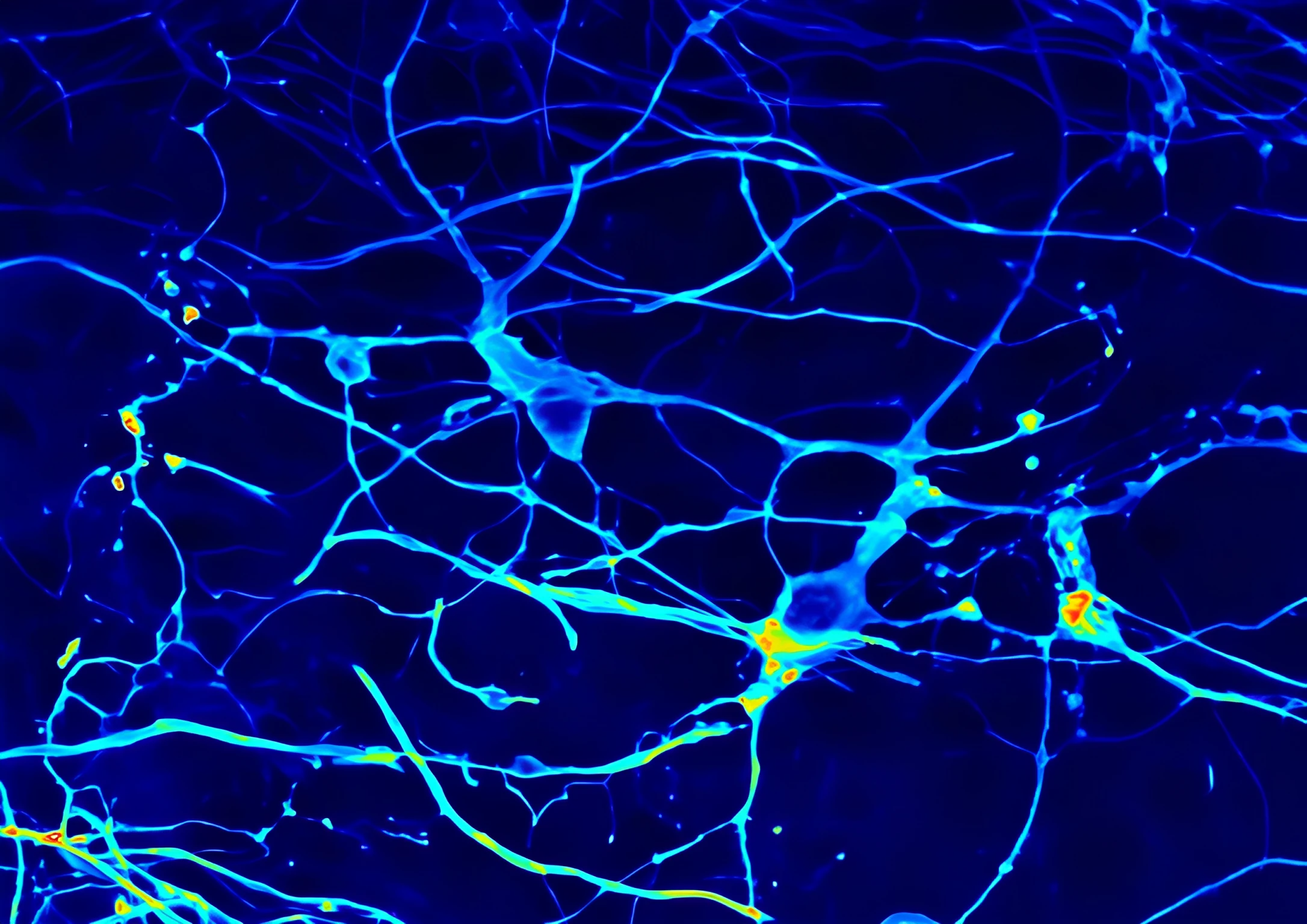
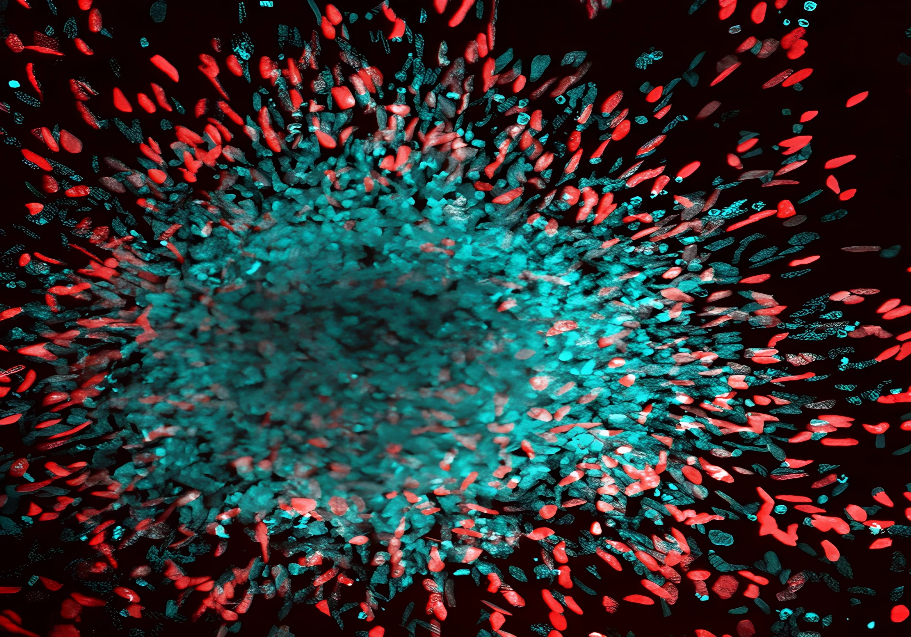
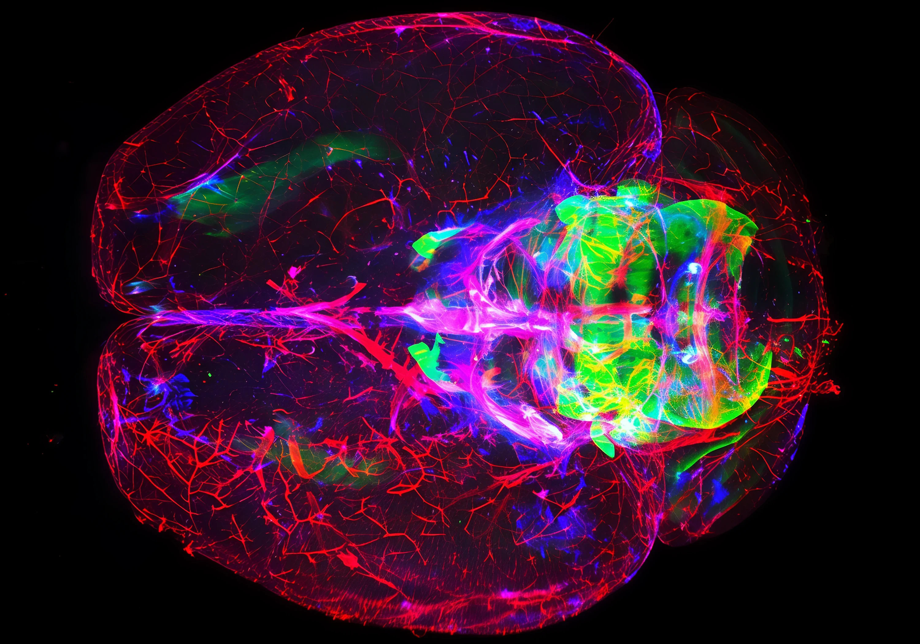
Glioblastome ; Institut du Cerveau
Yesim Guner
Yesim Guner plunges us into an in-depth view of a brain affected by incipient glioblastoma. Blood vessels form a dense network, while the first tumor cells silently infiltrate. A suspended moment, captured before the disease spreads.
Cancer du cerveau
Maladie d'alzheimer ; Pasteur
Claire Nivollet
Here, Claire Nivollet reveals a face of Alzheimer's disease: cyan clusters of β-amyloid proteins, encircled by the red extensions of astrocytes. In the face of invasion, these cells react, curl up and shake, between defense and disruption.
Maladie d'alzheimer
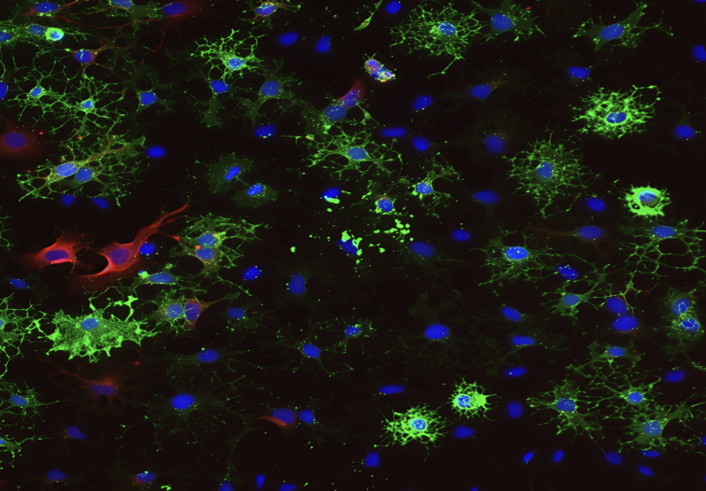
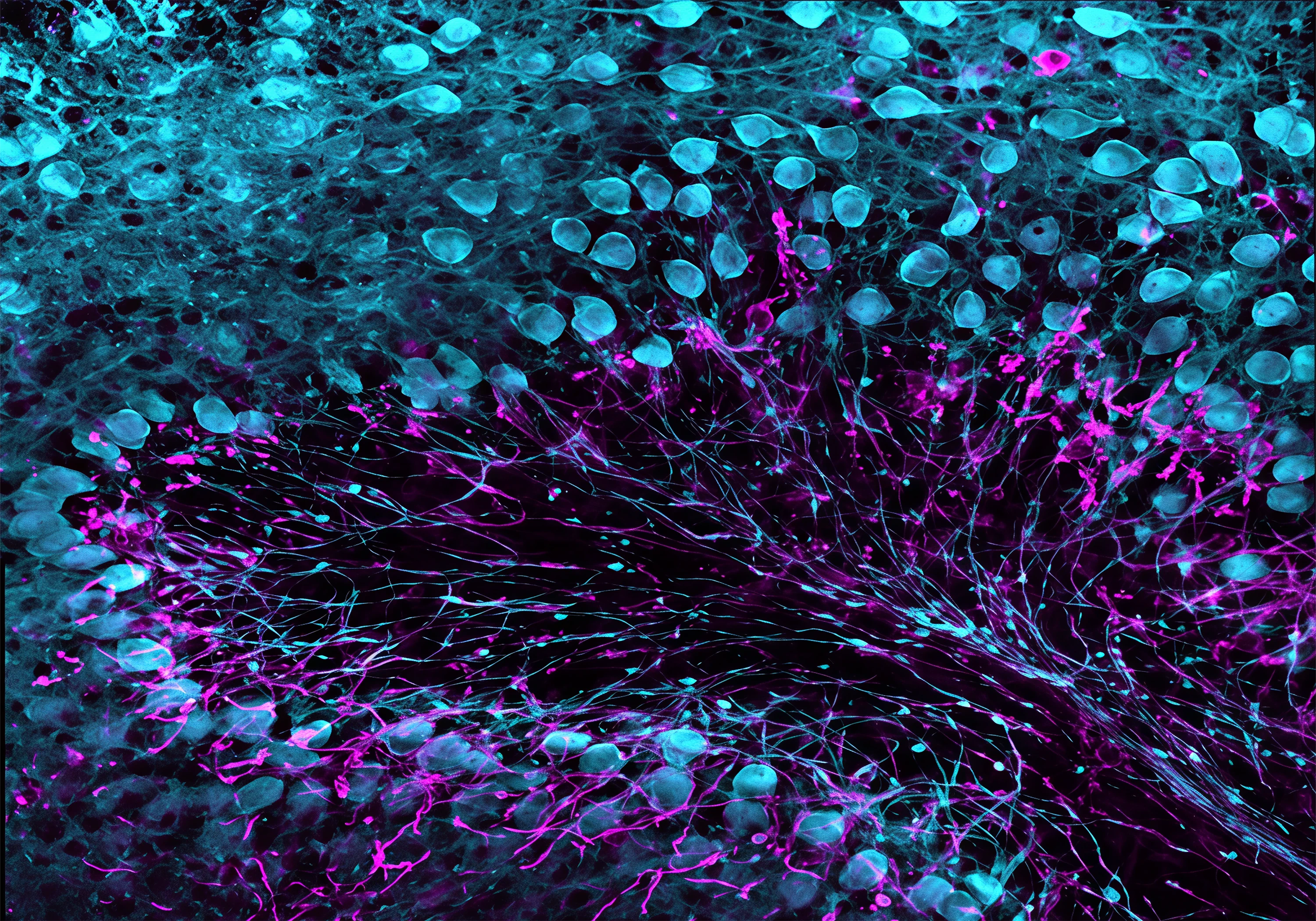
Maladie d'alzheimer ; Institut du Cerveau
Elodie Martin
Elodie Martin presents astrocytes, the brain's support cells, in a model of Alzheimer's disease. Their extensions form a dense meshwork, but in the pathology, their activity becomes dysregulated, amplifying inflammation and disrupting neuronal equilibrium.
Maladie d'alzheimer
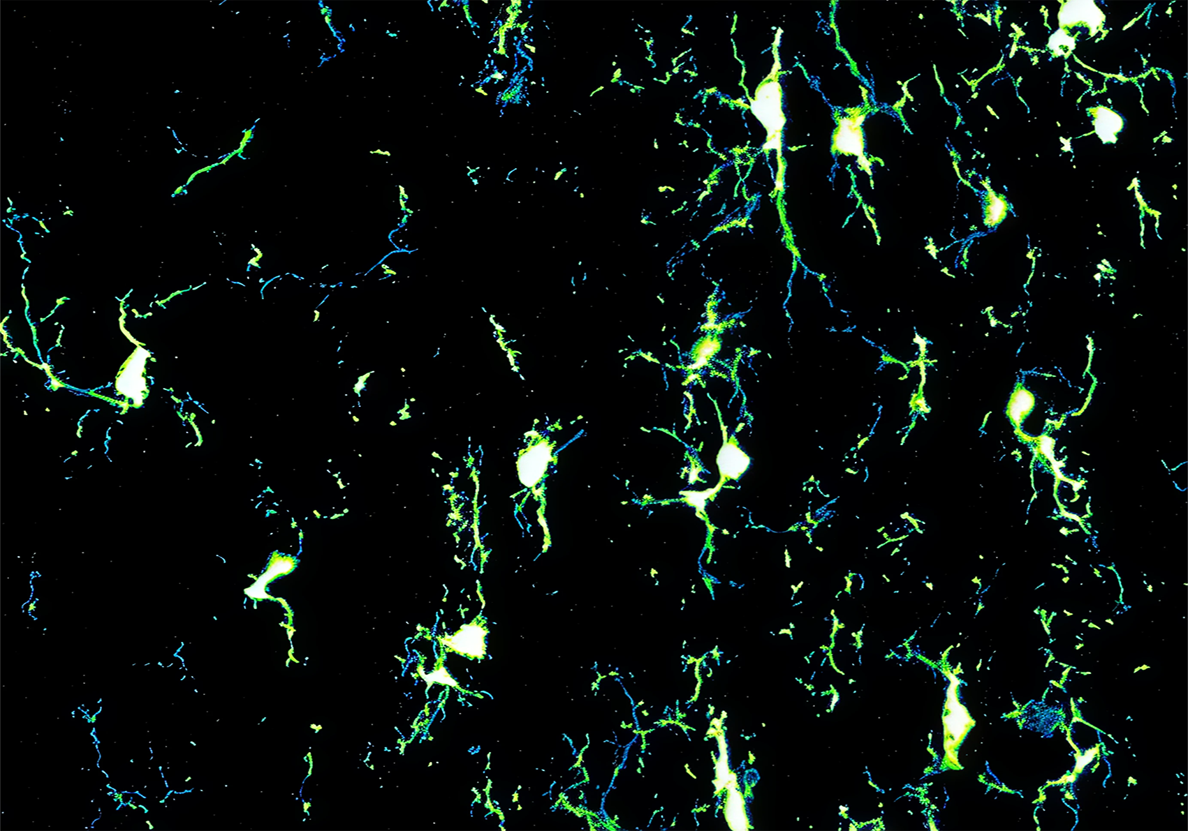
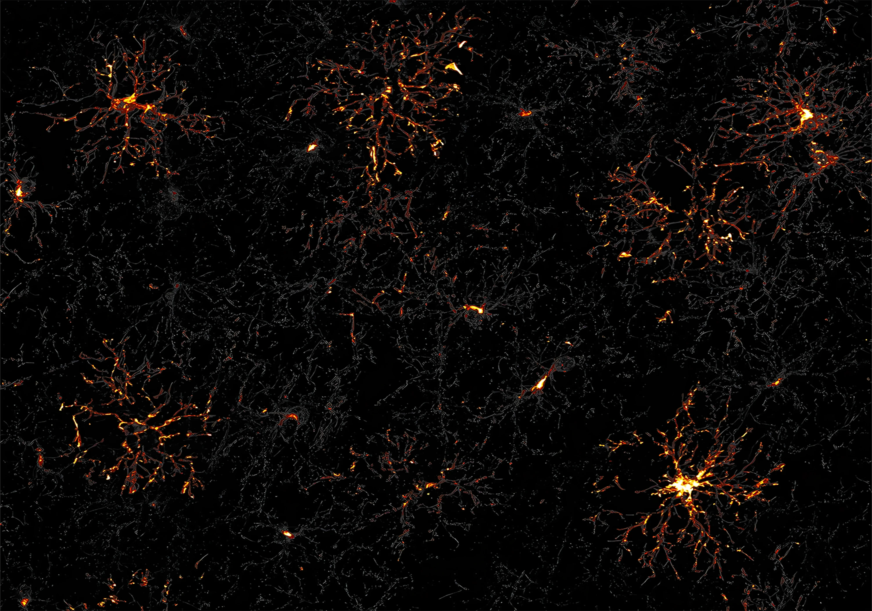
Réparation dans la sclérose en plaque ; Institut du Cerveau
Clément Perrot
Clément Perrot reveals microglia in the spinal cord, extending their fine extensions. Sentinels of the nervous system, these cells patrol relentlessly. At the slightest signal, they change shape, ready to repair, defend or disrupt.
Sclérose en plaque
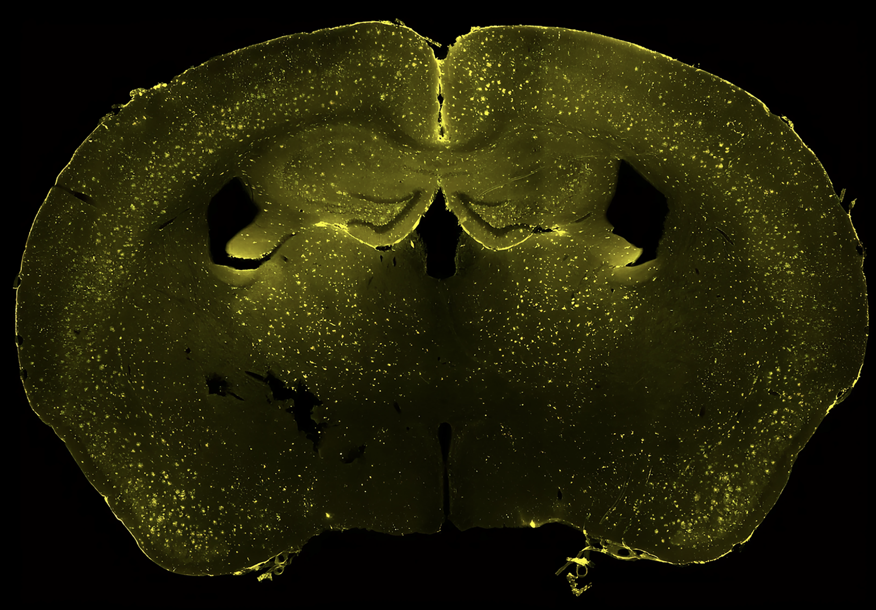
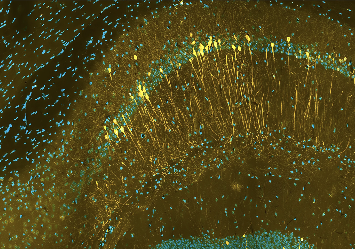
Maladie d'Alzheimer ; Institut du Cerveau
Jean David Randrianaly
Jean David Randrianaly invites you to observe the first invisible signs of Alzheimer's disease. In this cross-section of the brain, amyloid deposits glisten yellow, marking weakened regions long before the first signs of forgetfulness.
Maladie d'Alzheimer
Génétique et développement des tumeurs cérébrales ; Institut du Cerveau
Elisa Popa
Elisa Popa opens a window onto the silent dialogue between glial cells. In this image, you explore how the balance between support, signaling and inflammation can shift, long before the first visible signs.
Sclérose en Plaque
MIND : Métabolisme, Immunité et Neurodégénérescence ; Institut du Cerveau
Julie Pilon
Julie Pilon captures the hippocampus under construction. Neurons line up and weave their networks, sculpting the future circuits of memory. But an early imbalance can already be the beginning of a crisis.
Développement cérébral
Réparation dans la Sclérose en Plaque ; Institut du Cerveau
Tala Karam
Tala Karam maps the brain's major myelinized pathways. In green, myelin traces the networks of nerve impulses. Their silent alteration can disorganize circuits and hinder thought.
Slcérose en Plaque
Génétique et développement des tumeurs cérébrales ; Institut du Cerveau
Yvette Hayat
Yvette Hayat captures the disturbing cohabitation of neurons and tumor cells. In the face of glioma infiltration, circuits become disorganized, as if the cancer were silently redesigning the brain's language.
Cancer du Cerveau
Institut du Cerveau
Melika Ghaznavirad
Melika Ghaznavirad reveals the architecture of the cerebellum through Purkinje cells. Their dendritic branches, rigorously organized, orchestrate the balance and precision of gestures. Their alteration signals the first disagreements in movement.
Démence Frontotemporale
Réparation dans la Sclérose en Plaque ; Institut du Cerveau
Tala Karam
Tala Karam reveals a precise tangle of vessels, myelin and glial cells. This three-dimensional weave reveals the delicate balance between irrigation, signaling and repair, a balance that disease can silently disrupt.
Sclérose en plaque
Navigation, intégration sensori-motrice et interactions corps cerveau ; Institut du Cerveau
Claire Wyart
Claire Wyart reveals the first neurons in the process of differentiation, each marked by its own identity. But in certain genetic epilepsies, this spatial program is disrupted as early as the embryo, inscribing the seizure even before birth.
Développement cérébral
Navigation, intégration sensori-motrice et interactions corps cerveau ; Institut du Cerveau
Claire Wyart
Claire Wyart captures the spinal cord of a zebrafish embryo, illuminated by developmental genes. These signals trace the blueprints of the nervous system, where each cell chooses its future.
Développement cérébral
Institut NIAID
Anonymous
Produced by the NIAID Institute, this image shows HIV leaving a CD4+ T lymphocyte. Each emerging viral particle marks another step in the spread of the virus. A key moment, at the heart of strategies to halt the infection.
Virologie
Equipe "Tumour Microenvironment"
Hodivala-Dilke
K. Hodivala-Dilke captures a blood vessel infiltrating tumor tissue. A symbol of angiogenesis, this chaotic network nourishes the tumor as much as it betrays the imbalance: vital circulation diverted to the benefit of proliferation.
Cancer de la peau
University of California, Santa Barbara.
Gabriel Luna
Gabriel Luna reveals the neuronal complexity of the retina. Vessels and astrocytes work together to maintain eye balance. A tissue where each cell plays its own note... so that the brain can see.
Trouble de la vision
Jonathan Clarke
Jonathan Clarke reveals the pyramidal neurons, pillars of the cortex. Through their plasticity, they encode our memories. But in the face of Alzheimer's, epilepsy or stress, this architecture becomes fragile, a sensitive reflection of our memory in the making.
Mémoire, apprentissage
University of Genova
Yann Bernardinelli
Yann Bernardinelli reveals the astrocytes at the heart of the hippocampus. Far from being mere supporters, these glial cells orchestrate neuronal balance, participate in memory, and are altered in many brain pathologies.
Maladies Neurodégénératives
National Institutes of Health
Alex Ritter
Alex Ritter reveals actin bundles, the essential wiring of the cytoskeleton. Engines of movement, alignment and mechanical perception, these structures are altered in dystrophies, cancers and certain developmental disorders.
Cancer
BrainsRusDC
With their spectacular arborizations, Purkinje cells coordinate our gestures and fine-tune motor learning. As cerebellar sorting centers, their deterioration often signals the first signs of balance disorders.
Démence fronto-temporale
Institute of Child Health and Human Development
Dylan Burnette
Dylan Burnette reveals fibroblasts, the masters of the extracellular matrix. Support, healing, inflammation or cancer: these cells silently shape and transform the body's biological landscape.
Cancer
National Institute of Child Health and Human Development
Daniel Castranova
Daniel Castranova reveals the organization of the lymphatic system and skeleton in larval zebrafish. This co-visualization reveals the foundations of movement and immunity, on the scale of a developing organism.
Maladies Inflammatoires
École Polytechnique Fédérale de Lausanne
Blue Brain Project
A product of the Blue Brain Project, this virtual model simulates the electrical activity of neural networks. It paves the way for in silico exploration of memory, perception... and perhaps one day, consciousness.
Comportement et développement neuronal
Gull Lab
Samantha Griffiths
Samantha Griffiths captures trypanosomes in the midst of immune evasion. Masters of molecular disguise, these parasites melt into the bloodstream to infiltrate the body. A silent face-off between infection and defense.
Microbiologie
National Cancer Institute
Cecil Fox
Dr. Cecil Fox captures tumor cells in culture, figures of controlled disorder. Behind their autonomous proliferation lie mutations, disorders and potential points of action for better understanding and treating cancer.
Cancer
Masachusset Institute of Technology (MIT)
Kuan-Chung Su
Dividing LinesUnder the lens of Kuan-Chung Su, a cell in full division reveals the dysfunctions of cancer. Studying these stages of mitosis means understanding how proliferation becomes pathological, and how to stop it.
Cancer
Cell and Developmental Biology (MCDB)
Yi Zuo
Yi Zuo reveals the ramifications of the frontal cortex, where memory, learning and decision-making are sculpted. Each visible connection embodies a memory in the making, reflecting our capacity to think, adapt... or forget.
Mémoire, apprentissage
University College London
Ingrid Lekk and Steve Wilson
Ingrid Lekk and Steve Wilson capture the activation of a gene in the developing eye of a zebrafish embryo. A dive into the first stages of visual development, where life is written in light.
Maladie génétique et trouble du développement
///
Morne Arin
Morne Arni reveals the structure of Toxoplasma gondii, a parasite capable of entering, hiding and multiplying in our cells. Invisible in most people, it becomes formidable in fragile individuals or developing fetuses.
Lutte contre les infections parasitaires
Max Planck Institute for Human Cognitive and Brain Sciences
Alfred Anwander
Alfred Anwander unveils the connectome, the map of the brain's connections. Each bundle links key regions involved in memory, language or emotions. A precise vision of the brain in action... or under repair.
Compréhension du comportement humain
Réparation dans la Sclérose en Plaque ; Institut du Cerveau
Elodie Martin
Élodie Martin reveals the protective shadow of oligodendrocytes. In this image, you'll discover these cells that envelop neurons to accelerate nerve impulses. When they fail, as in multiple sclerosis, cerebral communication falters.
Sclérose en Plaque
Réparation dans la Sclérose en Plaque ; Institut du Cerveau
Clément Perrot
Clément Perrot reveals the architecture of the cerebellum: Purkinje cells and myelinated fibers orchestrate the precision of movement. A cellular symphony that falters in multiple sclerosis.
Sclérose en Plaque
Réparation dans la Sclérose en Plaque ; Institut du Cerveau
Noémie Frère
Noémie Frère reveals the harmony between astrocytes and neurons at the heart of brain tissue. This subtle interaction structures brain development - and falters in certain neurological disorders.
Développement cérébral / Autisme
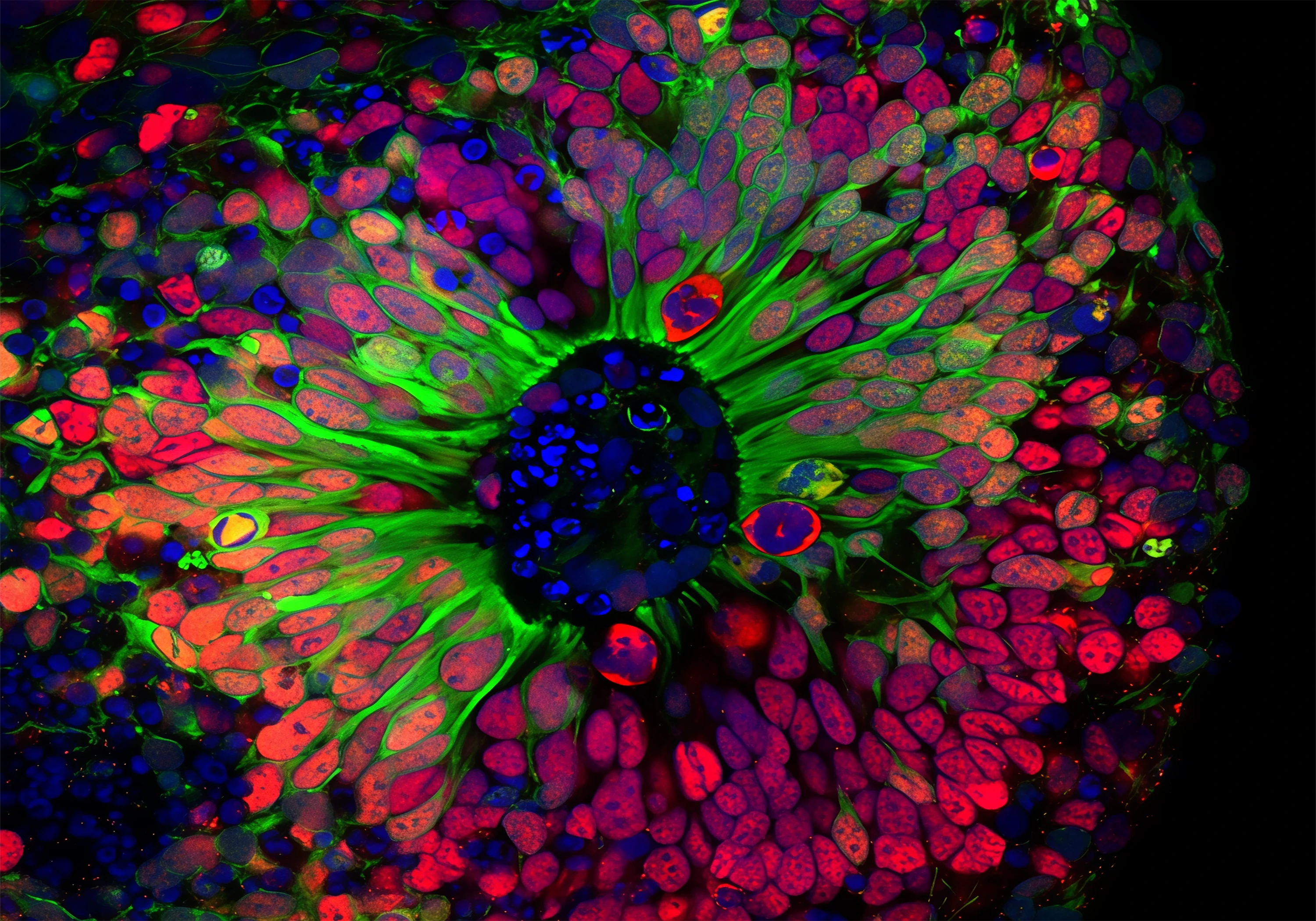
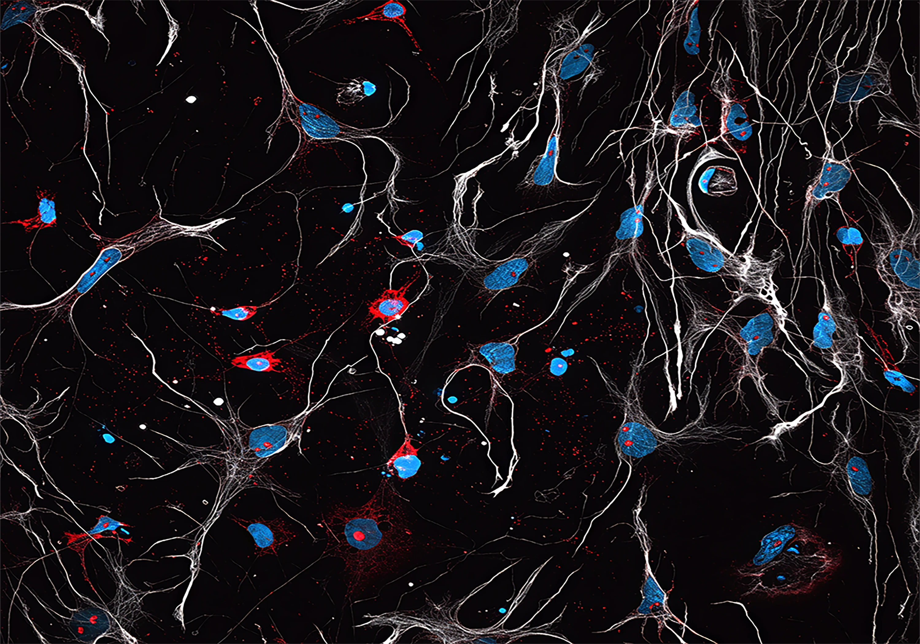
Génétique et physiopathologie de l'épilespise ; Institut du Cerveau
Marina Maletic
Marina Maletic explores a cerebral organoid derived from patient cells. This cohabitation of healthy and mutated cells reveals the invisible origins of certain forms of epilepsy, paving the way for personalized therapies.
Epilepsie
Développement et dysfonctionnement cérébral dans les maladies neurogénétiques; Institut du Cerveau
Melika GHAZNAVIRAD
Melika Ghaznavirad reveals the architecture of the cerebellum. Purkinje cells and granular cells organize themselves to orchestrate gesture. Their alteration lies at the heart of motor and neurodevelopmental disorders.
Démence frontotemporale / Maladie de Huntington